B0007605 Heartstrings
Credit: University of Oxford, Dr P.Hales/BBSRC. Wellcome Images
images@wellcome.ac.uk
http://wellcomeimages.org
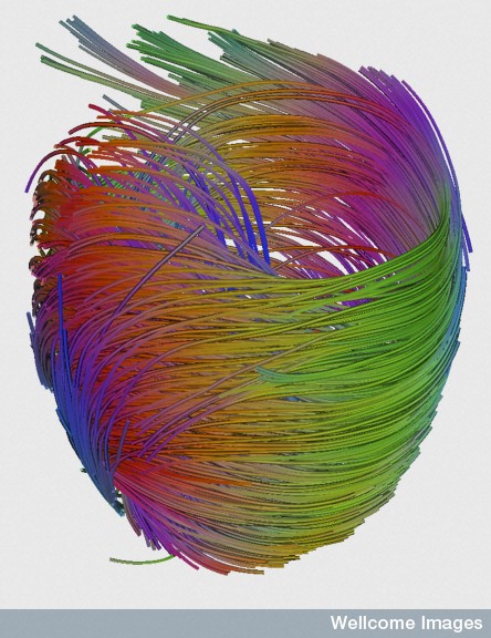
This image shows the swirling arrangement of cardiac fibres in the left ventricle. The myocardium (the heart’s muscular wall) is comprised of sheets of interconnected myocyte cells, which form these muscle fibres.
The heart consists of four chambers: the left and right atria and the left and right ventricles. The atria make up the upper region of the heart and the ventricles form the lower region. The left ventricle is responsible for pumping freshly oxygenated blood around the body and, therefore, has a thicker muscular wall compared to the right ventricle. The right ventricle is also imaged but is not observed here due to the viewing angle and region of interest chosen.
This image was produced using a branch of magnetic resonance imaging (MRI) called diffusion tensor imaging (DTI). The technique tracks the diffusion of water throughout the myocardium. Due to the orientation and organisation of the myocyte cells, the movement of water is restricted, so tracking the location of water molecules can reveal valuable information about the structure of the heart in a non-invasive way. This technology allows scientists to model the prevailing structure of muscle cells throughout the heart and how certain pathologies, such as ischemia, can cause this to change. Ischemia, otherwise known as angina, is a heart condition caused by reduced blood flow to the heart.
diffusion tensor imaging (DTI)
2010 Published: –
Copyrighted work available under Creative Commons by-nc-nd 4.0, see http://wellcomeimages.org/indexplus/page/Prices.html